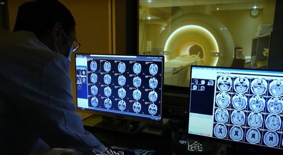
Magnetic Resonance Imaging (MRI Scan) is a non-invasive, radiation-free advanced diagnostic imaging technique that uses powerful magnetic fields and radio waves to generate high-resolution images of the inside of the body.
This scanning method is widely used across the body and is especially suitable for examining:
MRI is highly effective in helping medical professionals make accurate diagnoses and serves as an important tool for monitoring chronic conditions and health status changes.
It can detect hidden threats such as small tumors, vascular anomalies, and structural abnormalities, allowing timely medical or lifestyle intervention and improving treatment outcomes.
Regular MRI scan checkups help establish a personal health baseline, prevent disease progression, enhance wellness, and ultimately support longevity.
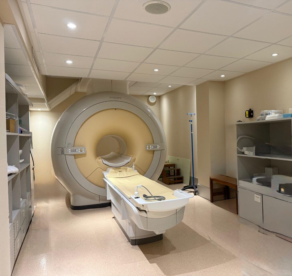
We offer a comprehensive MRI Service at the Magnetic Imaging Centre.